Transposing Bone Marrow Immune 'Neighborhoods' onto a Chip: How a 96-Well Platform Visualizes Antibody Cell Migration and Long-Term Survival
Original Chinese title: 把骨髓裡的免疫「鄰里」搬上晶片:96孔平台如何看見抗體細胞遷移與長期存活?
A 96-well bone marrow chip models perivascular and endosteal subniches to track the migration and survival of antibody-secreting cells; it is not complete human bone marrow, nor can it displace clinical trials or forecast individual immunity.
鄭淑禎|實踐大學專任助理教授
Focusing on industrial transformation, long-term care services, public service design, and technology applications, writing about how institutions genuinely ground themselves in users and frontline practice.
# Transposing Bone Marrow Immune 'Neighborhoods' onto a Chip: How a 96-Well Platform Visualizes Antibody Cell Migration and Long-Term Survival
It is widely established that following vaccination or natural infection, certain humoral immune responses persist for decades. Yet how antibody-secreting cells successfully navigate into human bone marrow, home to specialized niches, and sustain lifelong antibody production has historically defied direct observation. Situated deep within osseous cavities, bone marrow possesses an intricate microarchitecture of sinusoidal vasculature, hematopoietic lineages, stromal cells, and dynamic biochemical gradients. Conventional in vitro cell cultures can cultivate cells in flat dishes, but they fail to recreate spatial cues: who neighboring cells are, how interstitial fluids shear, and what structural microdomains mimic the perivascular niche. The transformative virtue of organ-on-a-chip engineering is converting these inaccessible microenvironments into observable, controllable experimental platforms.
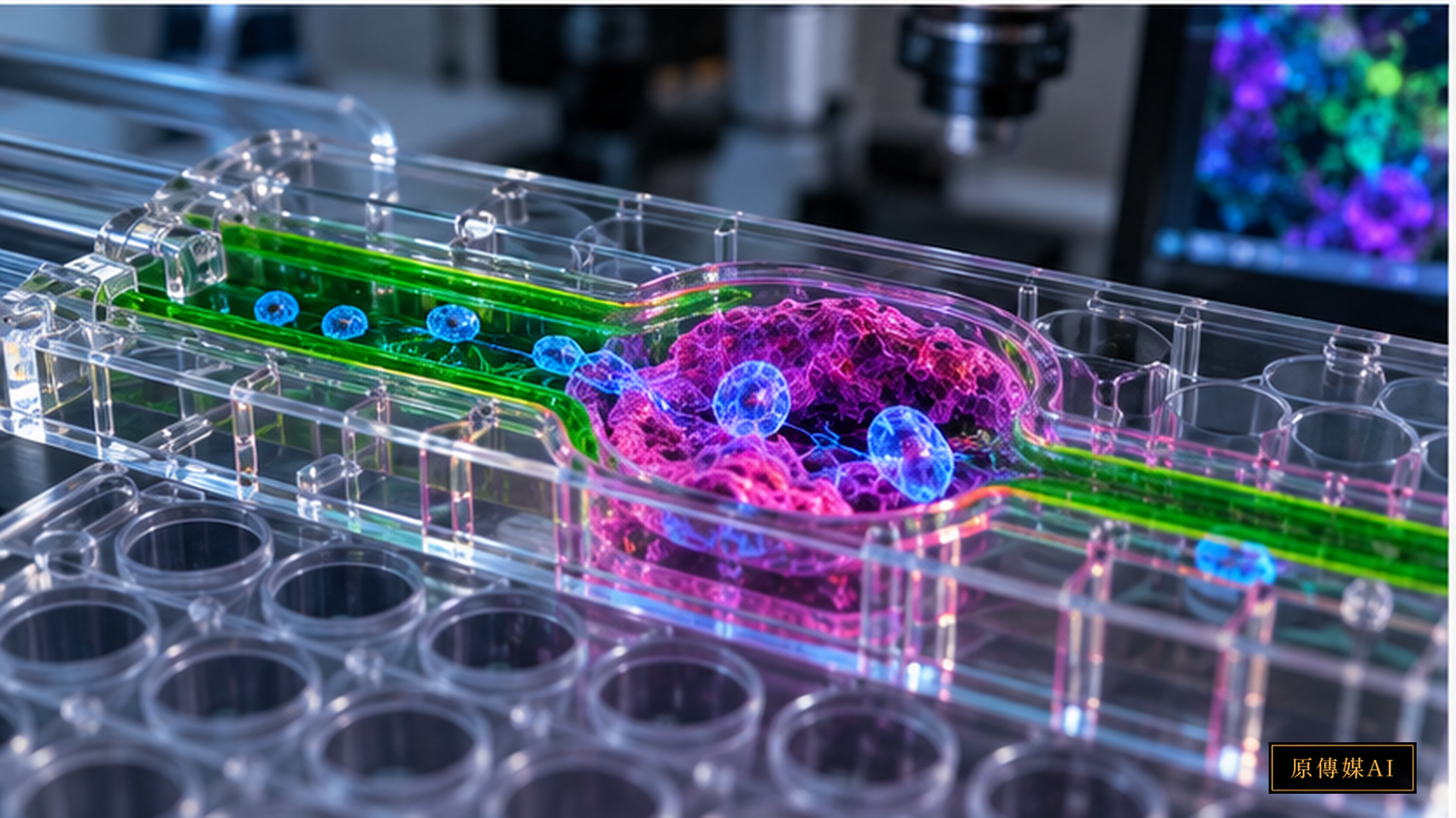
A human bone marrow-on-a-chip platform published in Science Advances in September 2026 integrates a perfusable microfluidic system with lymphoid tissue organoids, allowing investigators to track the continuous egress of immune cells from lymphoid tissues into simulated bone marrow compartments. The platform engineers distinct subniches mimicking perivascular and endosteal microenvironments, enabling high-content optical imaging within a standardized, higher-throughput 96-well plate format. Rather than terminating experiments at static timepoints to lyse cells for bulk assays, researchers can now directly visualize real-time cellular dynamics across space and time.
Investigators focused particularly on antibody-secreting cells (ASCs). Upon entering the chip, specific subsets preferentially migrated toward engineered perivascular zones, exhibiting a distinct "stop-and-go" motility pattern. These observations corroborate the paradigm that the bone marrow is not an undifferentiated cellular slurry, but a structured metropolis organized into distinct neighborhoods: specialized zones emit localized biochemical cues that arrest migration and sustain cell longevity. This demonstrates why cellular identity and physical spatial location must be investigated as an indivisible unit.
In conventional two-dimensional Petri dishes, cells adhere to rigid planar plastic, exposed to fluid mechanics and mechanical stiffness alien to human physiology. The bone marrow chip incorporates microfluidic perfusion, 3D extracellular matrix hydrogels, and organized co-cultures to construct a more physiologically relevant microenvironment. It remains, of course, a model: it lacks complete autonomic innervation, systemic hormonal axes, full immune cell repertoires, and multi-organ crosstalk. Nonetheless, it permits researchers to interrogate spatial and fluidic hypotheses far beyond the capabilities of planar culture.
This platform holds enduring promise for vaccinology. The clinical efficacy of a vaccine depends not merely on acute serum antibody titers, but on the successful generation and long-term maintenance of bone marrow-resident long-lived plasma cells. Benchmarking diverse adjuvant signals, donor cell backgrounds, or therapeutic candidates within this chip architecture allows scientists to dissect the conditions that favor durable humoral immunity. Nevertheless, it remains a laboratory discovery tool; it cannot forecast individual immune persistence following vaccination.
The implications extend equally to autoimmune disorders, allergies, and cellular senescence. Several autoimmune pathologies are driven by pathogenic plasma cells that find refuge within protective marrow niches, resisting immunosuppressive therapies. The chip provides a controlled testbed to evaluate how perturbing specific survival signals alters cell persistence. It accelerates hypothesis testing and mechanistic screening, though translating findings into clinical therapeutics still demands rigorous in vivo trials and human patient data.
The National Center for Advancing Translational Sciences (NCATS) at NIH has long championed tissue chips to bridge the translational valley of death, where candidate therapeutics succeed in animal models but fail catastrophically in human clinical trials. Human tissue chips integrate human cells into biomimetic microenvironments to generate predictive human data prior to clinical trials. Introducing bone marrow into this testing pipeline equips immunology and hematology with standardized, reproducible, and optically accessible translational models.
Rigorous science communication demands discipline here. Popular media readily brands organ chips as "synthetic humans" or declares that "clinical trials are obsolete"—claims that are scientifically groundless. Chips cannot displace human clinical trials. Their genuine value lies in rendering previously unobservable biological mechanisms quantifiable. They dramatically elevate experimental resolution, but they cannot replicate a patient's genetic polymorphism, clinical history, chronological age, polypharmacy, cumulative infections, or complex systemic physiology.
This is precisely where Two-Eyed Seeing bridges experimental models and clinical reality. Bioengineers optimize fluid shear stress, channel dimensions, and oxygen gradients; immunologists identify diagnostic cytokine signatures and phenotypic markers; clinicians navigate the messy heterogeneity of patient outcomes; and patients understand how diseases and treatment regimens alter their daily lives. If research pursues striking confocal microscopy without connecting to clinical dilemmas, chips become expensive technical novelties. Conversely, if clinical practice relies solely on broad diagnostic categories, it overlooks vital mechanistic clues.
From an educational perspective, this research offers a superb case study in the scientific method. Students learn that an experimental model is not a miniature replica of reality, but a deliberate abstraction where investigators isolate specific variables to answer defined questions. The best model is not the most baroque, but the one that knows precisely what it can and cannot resolve. This model literacy applies equally to artificial intelligence, climate forecasting, and public policy modeling.
Integrating bone marrow chips with single-cell transcriptomics, real-time intravital imaging, proteomics, and patient-derived primary cells will map the immune microenvironment with unprecedented fidelity. Yet the ultimate milestone is not replacing the human body with plastic chips, but deploying biomimetic platforms to eliminate false hypotheses early, identify robust therapeutic targets, and reduce translational drug failure.
Transposing bone marrow immune neighborhoods onto a microfluidic chip does not shrink human physiology into a 96-well plate; it grants science its first real-time window into how cells migrate, where they settle, and with whom they converse. Transforming invisible biological behaviors into quantifiable data expands our horizons, while reminding us that what we observe through the microscope is a model of reality—an invaluable stepping stone, but never the whole world.
A notable technological advance of this platform is the temporal dimension. Conventional endpoint assays only reveal cell counts or protein concentrations at fixed intervals; continuous live-cell microscopy captures whether cells halt, explore, or reverse direction. For immune cells, these kinetic patterns reveal niche interactions far more dynamically than static concentrations. Observing cells repeatedly arrest within specific microdomains allows researchers to systematically interrogate the adhesion molecules, chemokine gradients, or matrix stiffness governing that behavior.
Standardizing into a 96-well footprint aligns academic innovation with industrial drug screening. A multi-well microfluidic format allows investigators to evaluate dozens of variables concurrently: donor genotypes, therapeutic concentrations, matrix formulations, and cytokine cocktails. This converts piecemeal experiments into systematic comparative screens. However, high throughput demands stringent quality control: fluidic perfusion rates, cell seeding densities, and hydrogel polymer consistency across all 96 wells must remain tightly regulated to prevent technical artifacts from corrupting large datasets.
Regulatory science stands to benefit substantially. As organ chips establish analytical validity and reproducibility across independent laboratories, regulatory agencies like the FDA can incorporate chip data as qualified non-clinical evidence. Yet moving from academic proof-of-concept to formal regulatory qualification demands standardized operating procedures, cross-laboratory validation, certified reference materials, and defined contexts of use. Achieving academic publication is merely the first milestone; the harder work of translational science is ensuring protocols can be replicated reliably worldwide.
For patients, the true promise of this science is not an immediate miracle cure, but an expanded analytical toolkit to investigate why chronic illnesses persist and why patient responses diverge. Grounded, transparent communication builds lasting public trust. When sensationalized headlines promise instant breakthroughs, patients are misled; when scientists articulate exact capabilities and current limitations, genuine scientific literacy is nurtured.
Through the lens of Two-Eyed Seeing, patient narratives can inform early model design. If patients with autoimmune disorders struggle most with cyclical disease flares following therapy, engineers can prioritize modeling the persistence of refractory plasma cells; if specific demographics experience severe adverse reactions to immunotherapies, patient-derived cells can construct stratified niche models. In this way, organ chips transcend bioengineering showcases to directly address the questions patients need answered.
Reliable Sources
Role-Driven Inquiry
Yuan Media AI | Guided Questions by Role
Select a role first, then review the complete questions from that perspective; clicking a question will display Yuan Media AI’s response in real time.
Select a role
Immunology researcher
Examine the article's evidence, governance boundaries, data rights, and actionable steps from the perspective of a Immunology researcher.
Choose a question
AI use and content-safety disclosure
This article was compiled from official and research sources; established facts, research limitations, cultural contexts, and extended analyses are presented separately. The cover is an AI-assisted concept illustration.